M157
 4 pieces in 1 set
Size of 1 piece: H25 x W39 x D9cm
Weight (1 piece): About 2kg
Directors: Takaoka station south clinic president Kunio Tsukada, Kanazawa University premedical course Hiromi Sanada
![]()
![]()
The features and cautions of this model
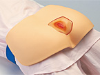
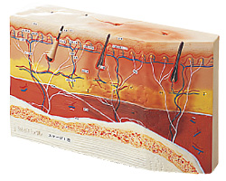
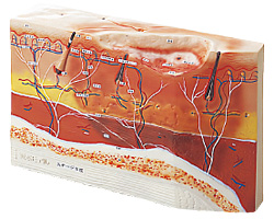
This Decubitus model was created with the hope that it will become a tool in understanding the eruption of Decubitus and the mechanism of its deterioration, so that one can choose the proper local treatment. When we do treatment on Decubitus, we can only see the surface. But what is the condition of the skin, hypodermal organ, muscles, and bones? We have made it possible to analyze the disease in its various stages from a sectional view, not only from observing the surface. By studying the conditions of Decubitus through this model, one can intuitively understand what is happening underneath the surface which should enable a more smooth choice of effective treatment.
Stage I
The epiderm has not yet been torn, but hypodermic bleeding and other red spots that do not disappear over a length of time are observed.
Stage 2
Abrasion, bubbles, bleeding, and effusion are observed.
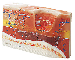
Stage 2I
Healing process of stage IV where the fascia and deeper had been affected.
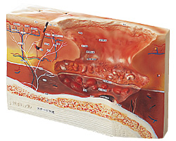
Stage IV
There is a hole widely separating the fascia and periosteum from the hypodermal organ.