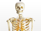
M170
 Weight: About 1kg
Case size: W92 x D21 x H18cm
Accessories: Storage case
![]()
![]()
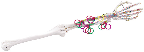
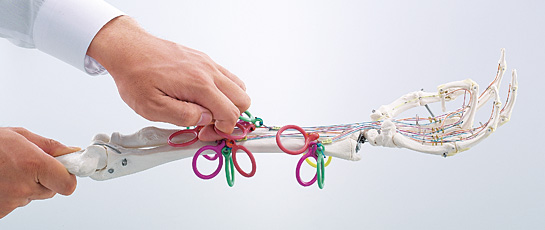
The mechanism of finger flexion can be visually checked during practice.
Distinguished by four colors of wires and rings, tendons engaged in the flexion of fingers can be visually identified and actually pulled, thus helping understand the mechanism of finger flexion instinctively.
The real-size model allows the users to compare with their own fingers during practice.
Practice
Flexion of fingers
1. Palmaris longus muscle (Green ring)
Originates from the medial epicondyle of humerus, passes over the flexor retinaculum, and spreads out in a fan-like form as the palmar aponeurosis. The palmaris longus muscle promotes skin tension. (Innervated by the median nerve)
2. Flexor digitorum superficialis muscle (Red ring)
Originates from the medial epicondyle of humerus, the ulna and the upper end of radius (radial head), and connects with the bottoms of middle phalanges of second to fifth fingers. (Innervated by the median nerve)
3. Flexor digitorum profundus muscle (Orange ring)
Originates from the upper front of ulna and the antebrachial interosseous membrane, separates into four tendons, and connects with the bottoms of distal phalanges of second to fifth fingers. The flexor digitorum profundus muscle bends the distal interphalangeal joints. (Innervated by the median nerve on the radial side and the ulnar nerve on the ulnar side)
4. Flexor hallucis longus muscle (Yellow ring)
Originates from the front of radius and the antebrachial interosseous membrane, and connects with the bottom of distal phalange of thumb. The flexor hallucis longus muscle bends the interphalangeal and metacarpophalangeal joints. (Innervated by the median nerve)